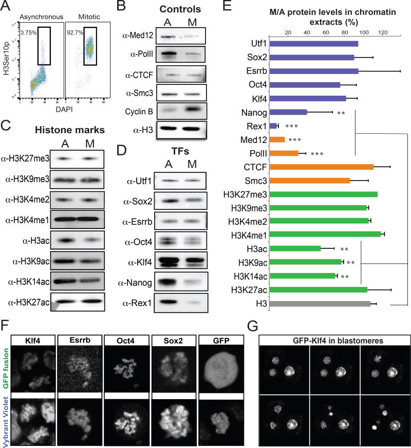
Figure 1. Histone modifications and pluripotency-related transcription factors are largely retained on the mitotic chromatin.
A. Representative FACS plots showing the percentage of asynchronous or nocodazole-treated ESCs (after mitotic shake-off) expressing the mitotic marker H3Ser10p. B–D. Western blot analyses showing the relative levels of positive and negative controls (B), selected histone modifications (C) and transcription factors (D) on the chromatin fraction of asynchronous (A) or mitotic (M) ESCs. E. Quantitation of (B–D) gel bands using Image J. The relative levels of each protein in mitotic chromatin fractions are plotted as a percentage of the respective levels in asynchronous chromatin fractions. Error bars indicate standard deviation based on at least two independent synchronization and fractionation experiments in two different ESC lines (ESC V6.5 and ZHBTc4.1). Asterisks indicate significant downregulation when compared to H3 levels, as calculated by t-test (**p<0.01, ***p<0.001). F. Representative live-imaging photos of mitotic ESCs expressing ectopic KLF4, ESRRB, OCT4 and SOX2 fused with a GFP reporter. Overexpression of GFP alone is used as a control. The chromosomes were stained using a cell-permeable DNA dye (Vybrant Violet). G. Time-lapse images of dividing blastomeres after microinjection of KLF4-GFP mRNA into one out of two blastomeres in 2-cell stage embryos (See also Supplemental Movie 1).

