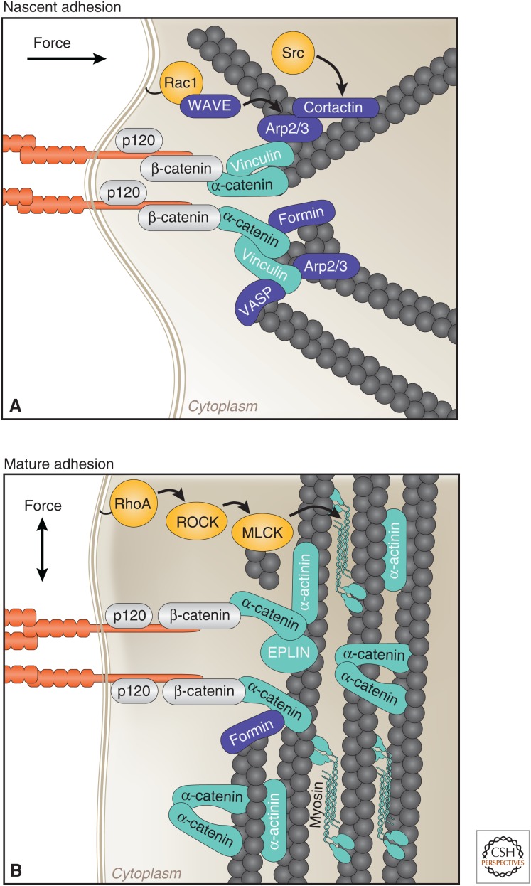
Figure 3.
Stages of cell–cell contact formation coincide with distinct organizations of the actin cytoskeleton, molecular compositions, and directions of forces. (A) Nascent cell–cell adhesions are connected to radial actin bundles lying perpendicular to sites of contact. These sites contain high levels of active Rac1 and Src, and actin-regulatory proteins (Arp2/3, cortactin, VASP, formin-1) that cooperate to generate a branched actin network required for contact expansion. α-catenin is under tension, which results in association with actin, directly through a catch–bond interaction, and vinculin. (B) At mature adhesions, the peri-junctional actin cytoskeleton is rearranged into bundles parallel to the plasma membrane. RhoA activity increases, which activates myosin II, thereby generating tensile forces required for adhesion maturation. Locally high concentrations of α-catenin presumably recruit actin binding and bundling proteins (EPLIN and α-actinin), and α-catenin itself forms homodimers that bind and bundle actin. EPLIN, epithelial protein lost in neoplasm (LIMA1); MLCK, myosin light-chain kinase; VASP, vasodilator-stimulated phosphoprotein.

