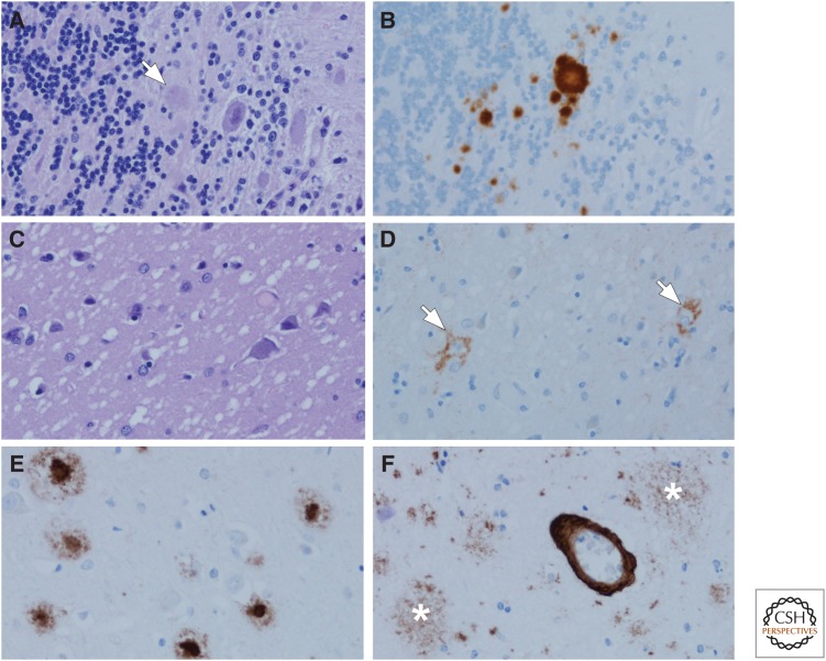
Figure 1.
Amyloidoses: Gerstmann–Sträussler–Scheinker disease (GSS) (A,B), Creutzfeldt–Jakob disease (CJD) (C,D), and Alzheimer’s disease (AD) (E,F). In GSS, dense-cored amyloid plaques can be detected with hemotoxylin and eosin (H&E) staining, arrow (A), but immunohistochemistry for human PrP (monoclonal antibody 3F4) reveals more clearly the multicentric nature of the deposits (B). One of the hallmark histologic features of CJD is the spongiform change in affected cortical and subcortical areas (C) with a perineuronal synaptic pattern of PrP deposition (arrows) in an adjacent section (D). In AD, amyloid deposits detected with immunohistochemistry (monoclonal antibody 6F/3D) are heterogeneous and include those with dense cores, especially in primary cortices (E), as well as poorly circumscribed and noncompact diffuse plaques in the cortex (asterisks) (F). In addition to parenchymal deposits, most cases of AD also have amyloid angiopathy (F).

