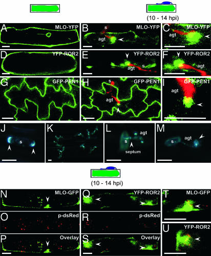
Fig. 1.
Induced polarity in host cells and powdery mildew sporelings. (A–I) Pathogen-triggered cell polarity in plant cells. A barley epidermal cell expressing MLO-YFP (A–C) or YFP-ROR2 (D–F) in the absence and presence of Bgh sporelings (10–14 hpi). (G–I) Nonchallenged and Bgh-challenged Arabidopsis epidermal cells expressing GFP-PEN1. Epiphytic fungal structures (red) were stained with propidium iodide. Stages of fungal development on cross-sectioned epidermal cells are depicted schematically above the columns. Images shown are single focal planes. Close-up views of attempted fungal entry sites for MLO-YFP, YFP-ROR2, and GFP-PEN1 expressing cells are shown in C, F, and I, respectively. White arrowheads mark the positions of focally accumulated fusion proteins. s, conidiospore. (Scale bar, 20 μm.) (J–M) Filipin-mediated fluorescence in Bgh conidiospores and at pathogen entry sites. Bgh-challenged leaf sections were stained with filipin. A representative conidiospore before germination (J), overview of germinated conidia (K) and a close-up view of germinated conidiospores (L and M) are shown. In J and L, arrowheads mark opposite poles of the conidium and the septum between the spore body and AGT, respectively. Note the filipin-mediated fluorescence underneath the appressorium in the attacked host epidermal cell (M, marked by arrowhead). s, conidiospore. (Scale bar, 20 μm.) (N–U) A barley epidermal cell expressing either MLO-YFP or YFP-ROR2 together with HvADF3 S6A blocking actin cytoskeleton function. Disruption of actin cytoskeleton was monitored by coexpression of a peroxisome-targeted dsRed (p-dsRed) variant in the same cells. (N–P) Coexpression of MLO-GFP, p-dsRed, and HvADF3 S6A. (Q–S) Coexpression of YFP-ROR2, p-dsRed, and HvADF3. (T and U) Close-up views of attempted fungal entry sites in the same cells. The stage of fungal development on a cross-sectioned epidermal cell is depicted schematically above the columns. (Scale bar, 20 μm.)

