Fig. 2.
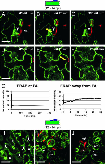
Focal PEN1 accumulation is triggered once and occurs at the cytoplasmic face of biotic stress sites. (A–F) Arabidopsis leaves expressing GFP-PEN1 were inoculated with Bgh conidiospores. At 12 hpi, FRAP analysis was performed at (A–C) and away from (D–F) focal accumulation (FA) sites. White arrowheads mark the position of focally accumulated GFP-PEN1 and the yellow flash depicts the site of photobleaching. (G) Quantitative measure of fluorescence recovery over time at and away from FA sites. Note that the fluorescence recovery is incomplete (≈75%) away from FA sites because of continuous photobleaching during imaging. (H–J) Bgh-challenged Arabidopsis leaves stably expressing GFP-PEN1 were imaged at 12 hpi before (H) and after (I and J) plasmolysis. st, stomata; he, Hechtian threads. The stage of fungal development on a cross-sectioned epidermal cell is schematically depicted above the columns. (Scale bar, 20 μm.)

