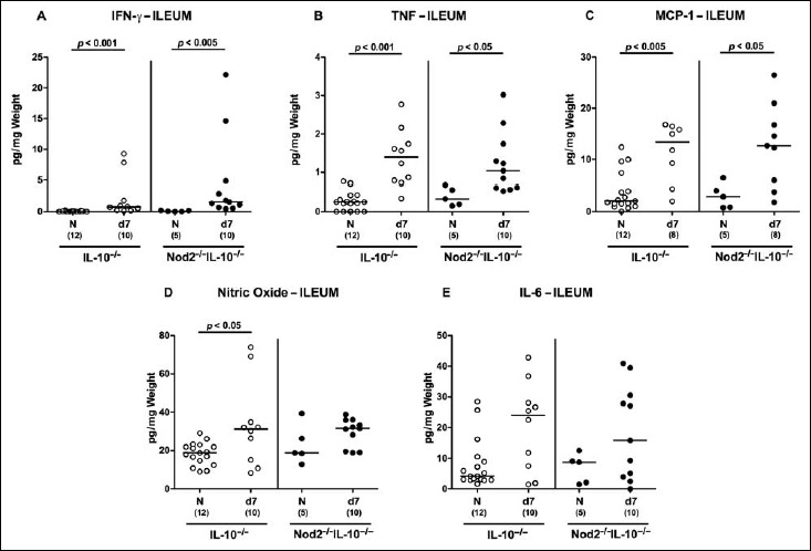
Fig. 6.

Ileal secretion of pro-inflammatory cytokines in C. jejuni strain 81-176 infected secondary abiotic IL-10–/– mice lacking Nod2. Secondary abiotic IL-10–/– (white circles) and IL-10–/– mice lacking Nod2 (Nod2–/– IL-10–/–; black circles) were generated by broad-spectrum antibiotic treatment and perorally infected with C. jejuni strain 81-176 by gavage at day (d) 0 and d1. (A) IFN-γ, (B) TNF, (C) MCP-1, (D) nitric oxide, and (E) IL-6 concentrations were determined in supernatants of ileal ex vivo biopsies at d7 postinfection. Naive (N) mice served as uninfected controls. Medians (black bars), level of significance (p value) determined by Mann–Whitney U test, and numbers of analyzed animals (in parentheses) are indicated. Data were pooled from two independent experiments
