Fig. 4.
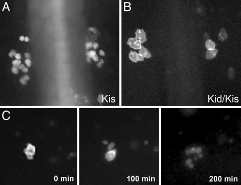
PGCs in kid-nos1 3′UTR treated embryos exhibit abnormal cell morphology culminating in cell death. Fluorescently labeled germ cells in kis-glo UTR (A) and kid-nos1 3′UTR/kis-glo UTR-injected (B) embryos at 12 hpf are shown. More PGCs are found in the control embryos and these appear smaller in size relative to kid-treated PGCs. (C) Frames from a time-lapse movie of a kid-nos1 3′-UTR-treated embryo showing a PGC undergoing cell death (see Movie 2).

