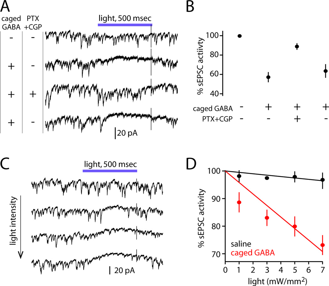
Figure 7. Evaluating GABA sensitivity using flash photolysis of caged GABA.
(A) Full-field photolysis of DPNI-caged GABA (390 nm light, 7 mW/mm2) transiently suppresses sEPSCs in a PN. Suppression is reversibly blocked by GABA receptor antagonists (5 µM picrotoxin, 50 µM CGP54626).
(B) Suppression requires caged GABA and functional GABA receptors (n=3, mean ± SEM). Light was delivered at 7–8 mW/mm2.
(C) In the presence of caged GABA, sEPSCs are increasingly suppressed by higher light intensities.
(D) Percent sEPSC activity and fits are calculated as in Figure 4F. Data is shown from one representative cell in caged GABA and one in regular saline (mean ± SEM across trials). Across a test set of PNs, the slopes of the fitted lines were significantly different in saline versus caged GABA (n=5 and 7, p=10−4, data not shown).

