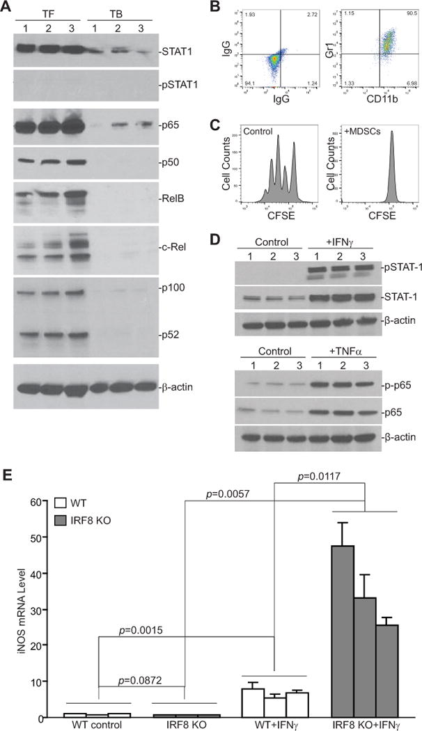
Figure 3. The IFNγ and NF-κB signaling pathways and MDSCs.

A. MDSCs were purified from the spleens of 4T1 tumor-bearing (TB, n=3) and their equivalent in tumor-free (TF, n=3) mice and analyzed by Western blotting for STAT1, pSTAT1, p65, p50, RelB, c-Rel, and p100/52. β-actin was used as a normalization control. B. 4T1 conditioned media was collected from 4T1 tumor cell culture flasks. BM cells from three WT mice were cultured in the presence of 4T1 conditioned media for 6 days. Cells were stained with IgG- or CD11b- and Gr1-specific mAb and analyzed by flow cytometry. Shown is the phenotype of the 4T1 conditioned media-induced MDSCs. C. CD3+ T cells were purified from the spleen of a tumor-free WT mouse and labeled with CFSE. The labeled T cells were then cultured in the absence or presence of 4T1 conditioned media-induced MDSCs at a 2:1 ratio for 3 days and analyzed for CFSE intensity by flow cytometry. Shown are representative data of proliferation of T cells from one of three replicates. D. BM cells from three WT mice were cultured in the presence of 4T1 conditioned media for 6 days. The resultant MDSCs were then either untreated (control) or treated with IFNγ (100 U/ml) or TNFα (100 U/ml) for approximately 20 hours, respectively. Cells were then analyzed by Western blotting for the indicated proteins. β-actin was used as a normalization control. E. BM cells from WT (n=3) and IRF8 KO (n=3) mice were cultured in the presence of 4T1 conditioned media for 6 days and then treated with IFNγ (100 U/ml) for approximately 20 hours. Cells were then analyzed by qPCR for iNOS mRNA level.
