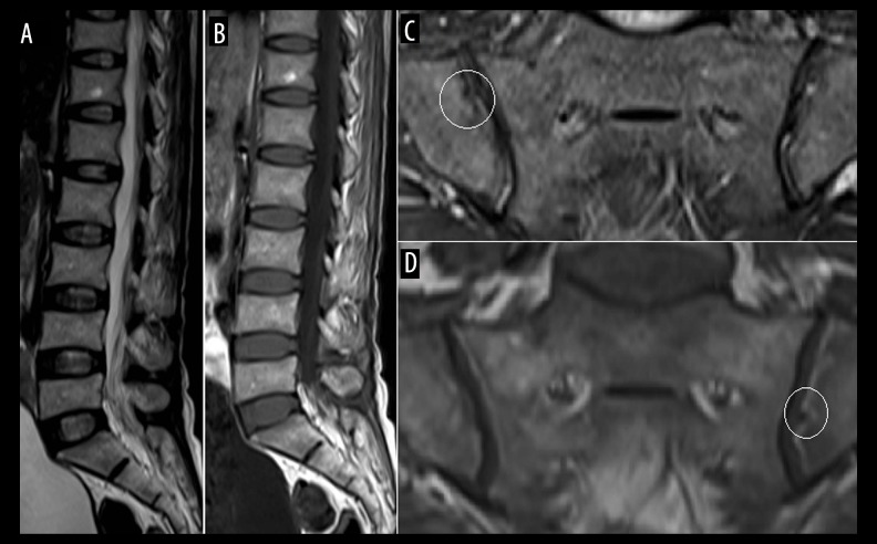
Figure 1.
(A) T2-weighted (W) and (B) T1-W sagittal MR images of the lumbosacral spine showing a mild decrease in height of the visualized thoracolumbar vertebrae; (C) T2 fat-suppressed oblique coronal and d, T1-W oblique coronal image of the sacroiliac joint showing mild irregularity along the iliac articular surface (circles), with no evidence of subchondral bone marrow oedema or significant erosion.

