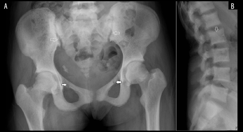
Figure 13.
(A) Frontal radiograph of the pelvis shows diffusely increased bone density, pseudo-fractures at the superior pubic rami (solid arrows); mild widening of the sacroiliac joint space and irregularity along the iliac articular surface (open arrow) suggest osteomalacia, note the bilateral ureteric calculi; (B) lateral radiographs of the lumbar spine show dense end-plates giving appearance of a rugger-jersey spine (open arrow). Renal osteodystrophy combines features of secondary hyperparathyroidism, osteomalacia and osteoporosis.

