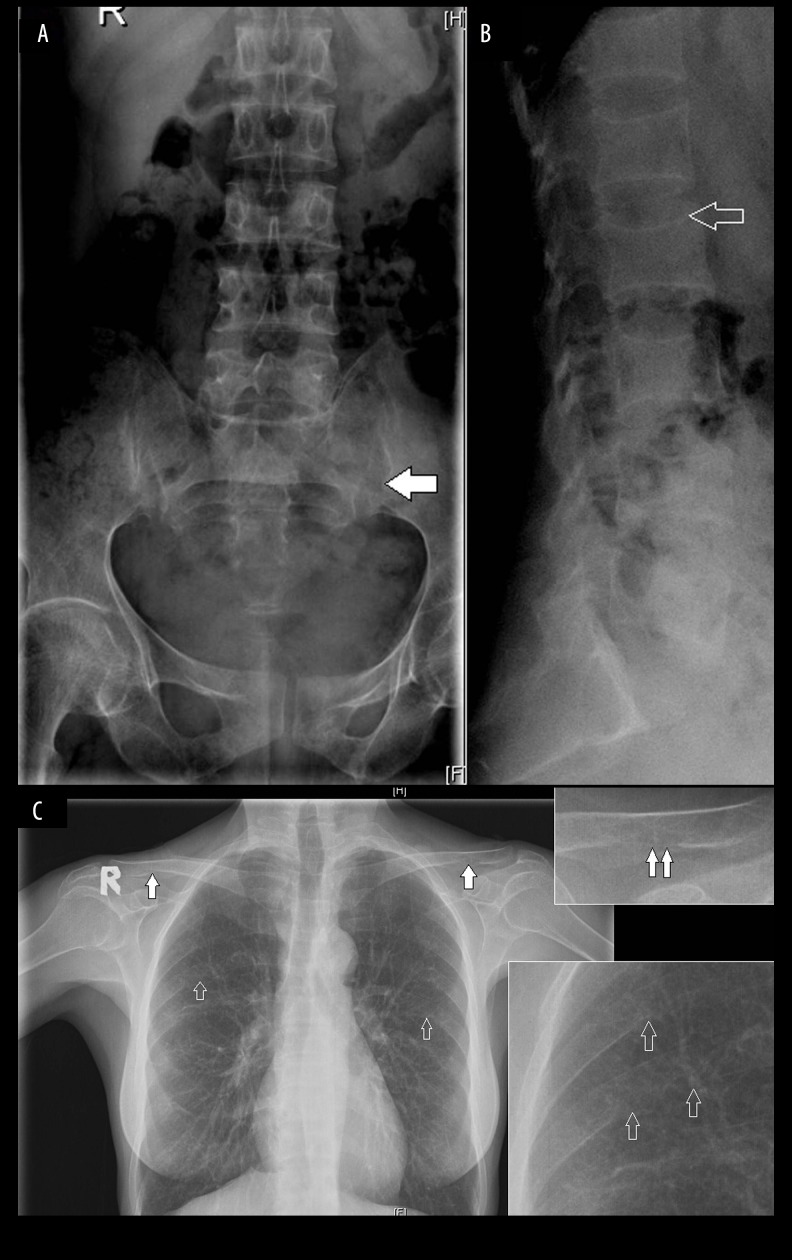
Figure 2.
(A, B) Frontal and lateral radiographs of the lumbosacral spine showing diffuse osteopenia of all the visualized bones with fuzzy bone density, widening of the sacroiliac joint with irregularity of the cortical joint line suggesting subchondral resorption along the iliac side (solid arrow). Multilevel decrease in height of the vertebrae with biconcavity and thinning of cortices (open arrow) suggesting bone softening; (C) Chest radiograph shows an indistinct margin with subperiosteal resorption along the inferior aspect of posterior ribs in the midclavicular line (open arrows), more appreciated in a magnified view. Similar subtle subligamentous resorption along the inferior surface of distal clavicle, more on the left side, clearly seen in a magnified view.

