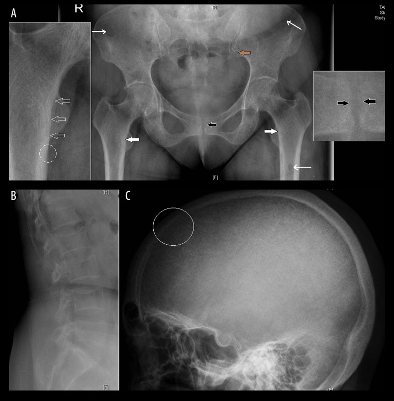
Figure 4.
(A) Frontal radiograph of the pelvis shows mild diffuse osteopenia, subtle subperiosteal resorption along the medial cortex of the upper femur bilaterally (solid white arrows), more clearly seen in the zoomed image, in the form of subtle periosteal cortical irregularity (open arrows) as compared to the adjacent normal periosteal cortex (circle); mild irregularity along the iliac side (brown arrow) of the sacroiliac joint suggests subchondral resorption; subchondral resorption along the pubic symphysis with a mild widening (black arrow) and multiple lytic lesions (thin long arrows) suggest brown tumour in the left femur and iliac bones; (B) Lateral radiograph of the lumbar spine and (C) skull show a decrease in height of the vertebrae with biconcavity and thinning of cortices; loss of definition of the inner table of skull (circle) with multiple tiny hyperlucent areas in the skull vault caused by resorption of the tubercular bone, giving a “pepper pot” appearance to the calvarium.

