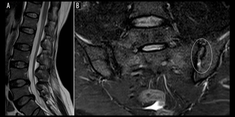
Figure 6.
(A) T2-W sagittal MR image of the lumbosacral spine shows a mild decrease in height of the visualized thoracolumbar vertebrae with mild disc disease in the lower lumbar spine; (B), T2 fat-suppressed oblique coronal view of the sacroiliac joint shows mild irregularity along the iliac articular surface with widening of joint space (circle).

