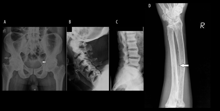
Figure 8.
(A) Frontal radiograph of the pelvis shows diffusely increased bone density, trabecular thickening, thickening and ossification of the sacrotuberous ligament (solid arrow), ossification of the rectus femoris tendon attachment at the anterior inferior iliac spine (open arrow); (B, C) Lateral radiographs of the cervical and lumbar spine show a diffuse increase in bone density, thickening and ossification of the anterior (open arrow) and posterior (black arrow) longitudinal ligaments; (D) frontal radiograph of the right forearm shows interosseous membrane ossification (arrow).

