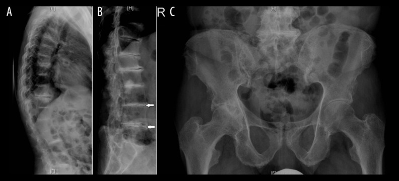
Figure 9.
(A, B) Lateral radiographs of the thoracic and lumbar spine demonstrate multilevel disc space narrowing, intervertebral disc calcification (arrows), vertebral osteophytes and mild osteopenia; (C) Frontal radiograph of the pelvis shows degenerative arthritis of the right hip joint and both sacroiliac joints.

