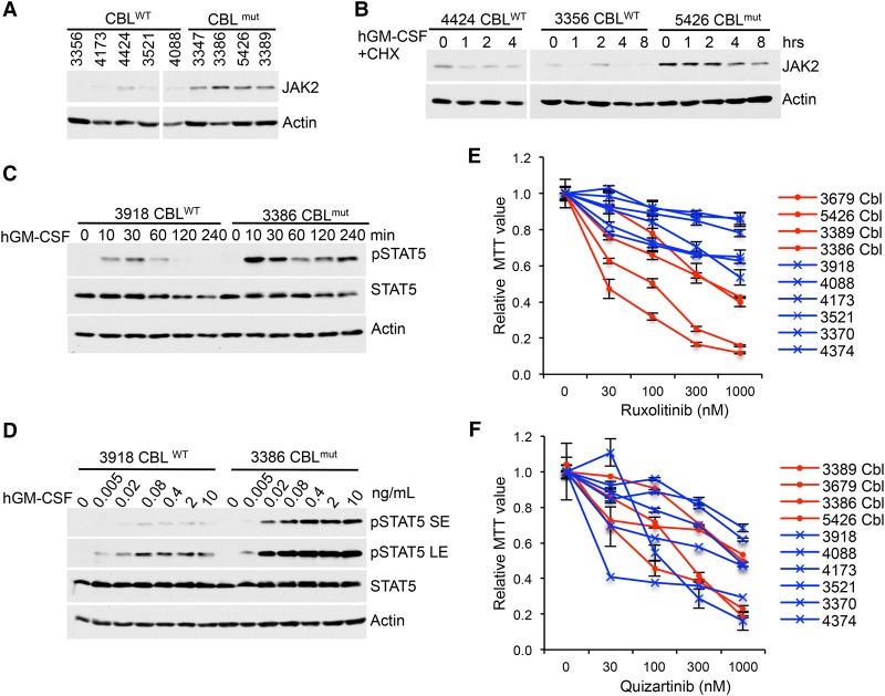
Figure 6.
CBLmut AMLs show enhanced STAT5 activation, elevated JAK2 levels, and hypersensitivity to JAKi compared with CBLWT AMLs. (A) Equal numbers of peripheral blood-derived MNCs and BM-derived MNCs from CBLWT and CBLmut AMLs were subjected to Western blot for JAK2 levels. (B) JAK2 half-lives in CBLWT and CBLmut AML cells were examined by Western blot upon GM-CSF and CHX treatment. (C,D) CBLWT and CBLmut AML cells were stimulated with GM-CSF for the indicated times (C) or with a graded dose of GM-CSF for 10 min (F). Cell lysates were subjected to Western blot analysis with the indicated antibodies. (E,F) AML cells were plated in cytokines and a graded dose of ruxolitinib (E) or quizartinib (F). Forty-eight hours later, live-cell numbers were quantified by MTT. CBLmut samples are labeled as “Cbl” in red lines, while CBLWT samples are indicated in blues lines.

