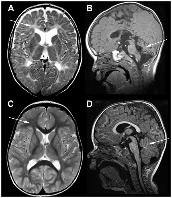
Fig. 6. MRI in the patient with TUBB4A-related severe hypomyelination and atrophy of the cerebellum.
A, B) T2-weighted axial (A) and T1-weighted sagittal (B) images of the patient obtained at the age of 2½ years; C, D, same images of an age-matched neurologically normal child. In the patient the cerebral white matter is T2-hyperintense due to lack of myelin (arrow in A), while the white matter in the control is T2-hypointense (arrow in C). In the patient the cerebellum is mildly atrophic (arrow in B), while it is normal in the control (arrow in D).

