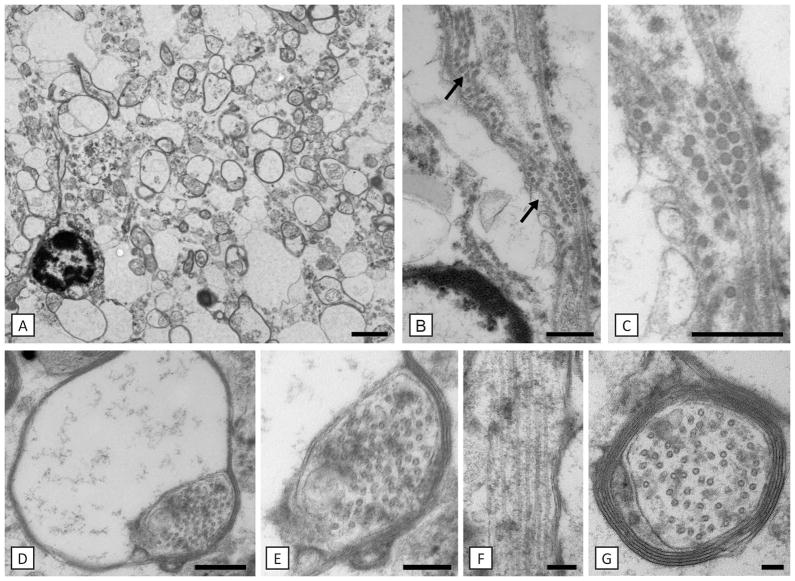
Fig. 8. Hypomyelination is associated with microtubule accumulation in the patient.
Axons are either non-myelinated or have thin myelin sheaths (A). Close to a putative oligodendrocyte nucleus there are rows of microtubules (arrows in B). Magnification of one of these areas is shown in C). The most distal part of the oligodendrocyte process, the inner loop is enlarged and filled with microtubules (D, E). An oligodendrocyte process seen on longitudinal section contains prominent microtubules (F). Microtubules were also prominent in myelinated and non-myelinated axons (G). Scale bars: 100 nm (G), 200 nm (E, F), 400 nm (C, D), 500 nm (B), 2 μm (A).

