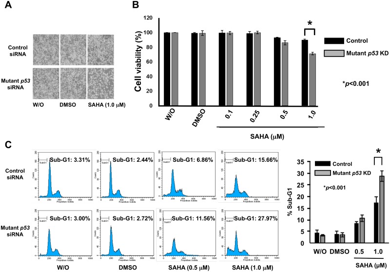
Fig 4. Silencing of mutant p53 in MiaPaCa-2 cells stimulates SAHA-dependent decrease and increase in cell viability and cell death, respectively.
(A) Phase-contrast micrographs. MiaPaCa-2 cells were transfected with control siRNA or with siRNA against p53, and then treated with DMSO, 1 μM of SAHA or left untreated. Forty-eight hours after treatment, the representative pictures were taken. (B) WST assay. MiaPaCa-2 cells were transfected with control siRNA or with siRNA against p53, and treated with DMSO or with the indicated concentrations of SAHA. Forty-eight hours after SAHA exposure, cells were analyzed by the standard WST cell survival assay. Solid and grey boxes indicate control siRNA- and p53 siRNA-transfected cells, respectively. (C) FACS analysis. MiaPaCa-2 cells were transfected with control siRNA or with siRNA against p53, and treated with DMSO or with the indicated concentrations of SAHA. Forty-eight hours after treatment, floating and adherent cells were harvested and subjected to flow cytometric analysis. Solid and grey boxes indicate control siRNA- and p53 siRNA-transfected cells, respectively.

