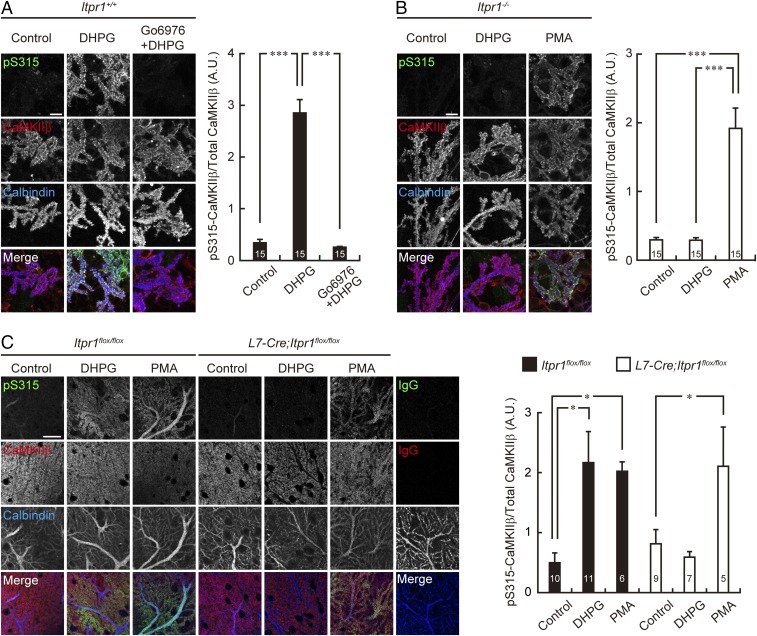
Fig. 3.
mGluR stimulation increases CaMKIIβ phosphorylation at S315 through IP3R1/PKC signaling. (A and B) Immunostaining of PKC-mediated phosphorylation of CaMKIIβ at S315 upon mGluR stimulation in cultured Itpr1+/+ and Itpr1−/− Purkinje cells. Primary cultured cerebellar cells from Itpr1+/+ mice were treated with 100 μM DHPG for 10 min in the presence or absence of 5 μM Go6976 (A), and cells from Itpr1−/− mice were treated with 100 μM DHPG or 0.4 μM PMA for 10 min (B). Treated cells were immunostained with the antibodies indicated. Purkinje cell distal dendritic areas are shown. (Scale bars, 10 μm.) Quantification of the phosphorylation level of CaMKIIβ at S315 in the distal dendritic area of Purkinje cells are shown at Right. ***P < 0.0001, one-way ANOVA with Bonferroni’s test for multiple comparisons. (C) Immunohistochemistry of S315 phosphorylation of CaMKIIβ of Purkinje cells in acute cerebellar slices prepared from Itpr1flox/flox and L7-Cre;Itpr1flox/flox mice. The slices were treated with 100 μM DHPG for 5 min or 0.4 μM PMA for 15 min, resectioned, and immunostained with the indicated antibodies. Distal dendritic areas of Purkinje cells are shown. (Scale bar, 20 μm.) Quantification of the phosphorylation level of CaMKIIβ at S315 in Purkinje cells is shown at Right. *P < 0.05, one-way ANOVA with Dunnett’s multiple-comparison post hoc test compared with control within each genotype. The numbers of neurons (A and B) and sections (C) are indicated in each graph.

