Fig. 1.
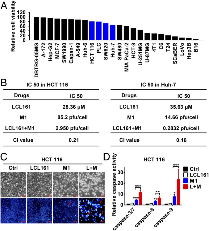
SMCs enhance M1-induced apoptosis in tumor cells. (A) Relative cell viability in 24 tumor cell lines treated with M1 [multiplicity of infection (MOI) = 1 plaque-forming unit per cell (pfu/cell), 48 h]. (B) IC50 and CI values in HCT 116 and Huh-7 cells. IC50 values are indicated in Fig. S1 A and D by vertical dotted lines, and the calculation formula for the CI is detailed in SI Materials and Methods. (C) Phase-contrast and Hoechst 33342 staining (5 μg/mL for 10 min) of HCT 116 cells treated with M1 (MOI = 1 pfu/cell) with or without 5 μM LCL161 for 72 h. (Scale bars: 50 μm.) (D) Relative activities of caspase-3/7, caspase-8, and caspase-9 were detected in HCT 116 cells treated as in C. Error bars represent mean ± SD obtained from three independent experiments. Ctrl, control; L+M, LCL161 + M1. *P < 0.05; **P < 0.01; ***P < 0.001.

