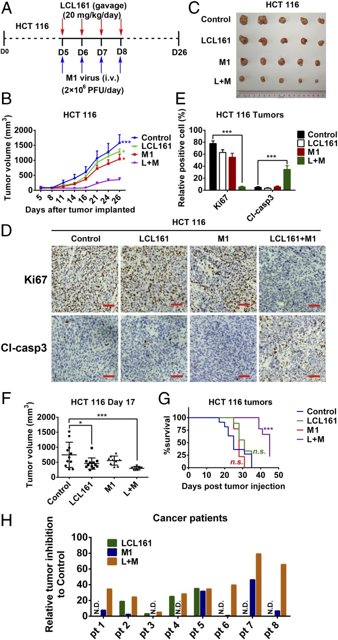
Fig. 6.
Combination of LCL161 and M1 inhibits tumor progression in a mouse xenograft model and in human ex vivo tissues. (A–C) Effect of LCL161 and M1 combination on the mouse xenograft model with HCT 116 tumors (n = 5, tumor volume in each group was compared with the control group). D, day. (D) Intratumoral expression of Ki67 and cleaved caspase-3 (Cl-casp3) was detected by immunological histological chemistry in HCT 116 tumors. (Scale bars: 50 μm.) (E) Quantitation of cells positive for Ki67 and Cl-casp3 from multiple HCT 116 tumors and relative positive cells are shown (n = 5). (F and G) HCT 116 cells were implanted in nude mice when the tumor volume reached about 150 mm3. Nude mice were treated with vehicle, LCL161, M1, and LCL161 plus M1 as in A. (G) According to the Animal Ethical and Welfare Committee of Sun Yat-sen University, when the tumor volume reached 1,500 mm3, the mice were euthanatized for the survival curve drawing. (F) Tumor volume reached 1,500 mm3 first in the control group at day 17 (control, n = 12; LCL161, n = 12; M1, n = 9; LCL161 + M1, n = 9.). (H) Surgical colon cancer specimens (50–100 mg) were cut into small pieces (∼1 mm3) and cultured in DMEM with 10% FBS and 5% penicillin/streptomycin at 37 °C. Tissues were then treated with vehicle (OPTI SFM), M1 (5 × 10⁷ pfu), LCL161 (10 μM), and LCL161 (10 μM) plus M1 (5 × 10⁷ pfu) for 3 d. Activity of tissues was measured by tissue culture end-point staining computer image analysis after staining with 3-(4,5-dimethylthiazol-2-yl)-2,5-diphenyltetrazolium bromide. The inhibition rates of each treatment for eight patients are shown. Error bars represent mean ± SD. pt, patient. *P < 0.05; ***P < 0.001.

