Fig. 2.
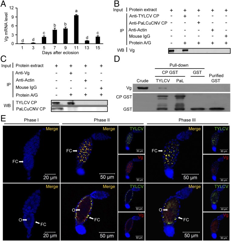
Vg is involved in TYLCV entry into whitefly oocyte. (A) The expression patterns of Vg in adult female whiteflies at different developmental stages. Mean ± SEM of three experiments. P < 0.05 (one-way ANOVA, LSD test). (B and C) Coimmunoprecipitation (CO-IP) of Vg with anti-CP monoclonal antibody (B) and vice versa (C) in whitefly crude extracts. (D) B. tabaci endogenous Vg coeluted with GST-fused TYLCV CP but not with GST-fused PaLCuCNV (PaL) CP. (E) Localization of TYLCV and Vg in follicular cells (upper lane) and oocytes (lower lane) of ovarioles at different developmental phases. Cell nucleus was stained with DAPI (blue). For phases II and III, both Vg (red) and CP (green) are shown. Yellow color indicates the overlay of red and green. FC, follicular cells; O, oocyte.

