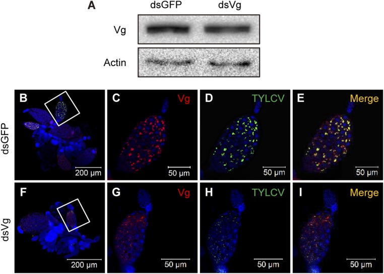
Fig. S3.
Knocking down the expression of Vg reduced yolk accumulation into oocytes and impeded entry of TYLCV into the ovary. (A) Vg protein levels after feeding with dsRNAs. (B–I) Localization of TYLCV and Vg in ovaries of dsGFP (B–E)- or dsVg (F–I)-treated whiteflies after a 48-h AAP on TYLCV-infected plants. C–E are the magnification of the boxed area in B, and G–I are the magnification of boxed area in F. TYLCV virions were detected by use of a rabbit anti-CP polyclonal antibody and goat anti-rabbit IgG labeled with Dylight 488 (green) secondary antibody. Vg was detected by use of a mouse anti-Vg monoclonal antibody and goat anti-mouse IgG labeled with Dylight 549 (red) secondary antibody. Cell nucleus was stained with DAPI (blue). Yellow color indicates the overlay of red and green.

