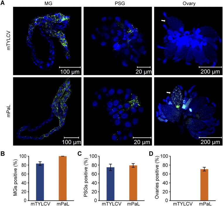
Fig. S5.
Localization of mTYLCV or mPaLCuCNV in different tissues of MEAM1 whitefly. (A) Localization of mutant TYLCV (mTYLCV) or mutant PaLCuCNV (mPaL) in midguts (MGs), primary salivary glands (PSGs), and ovaries of viruliferous whiteflies at 11 DAE. Mutant TYLCV (mTYLCV) and mutant PaLCuCNV (mPaL) virions were detected by use of a mouse anti-CP monoclonal antibody and goat anti-mouse IgG labeled with Dylight 488 (green) secondary antibody. Cell nucleus was stained with DAPI (blue). (B–D) The proportion of mutant TYLCV (mTYLCV) or mutant PaLCuCNV (mPaL)-positive MGs (n = 24) (B), PSGs (n = 24) (C), and ovaries (n = 24) (D). Mean ± SEM from three experiments. P < 0.05 (one-way ANOVA, LSD test). White arrows indicate mature oocytes.

