Fig. 1.
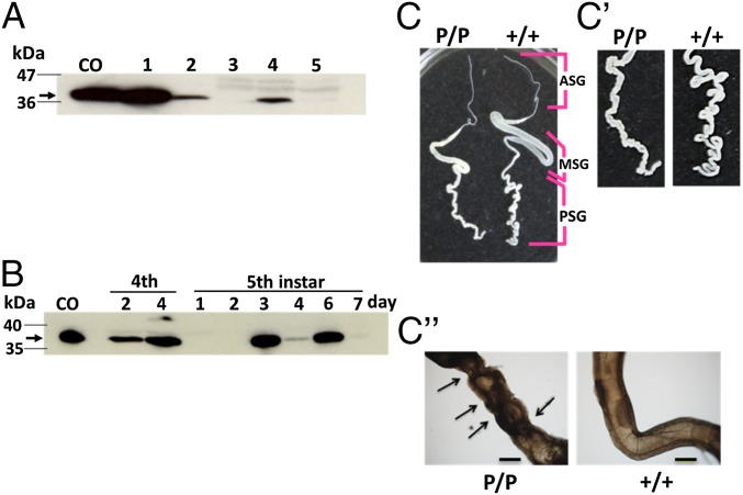
Expression of the truncated pierisin-1 protein (P1A269) in PSGs from w1-pndP1A269/P1A269 silkworms (B. mori). (A) P1A269 protein detection in PSGs by immunoblotting with a FLAG-tag antibody: lane CO, cypovirus polyhedron-encapsulated P1A269; lanes 1 and 2, P1A269-expressing Sf21 and BM-N cells, respectively (SI Appendix, Figs. S4 and S5); lane 3, PSGs from w1-pnd+/+ on day 7 of the fifth instar; and lanes 4 and 5, PSGs from w1-pndP1A269/P1A269 on days 6 and 7 of the fifth instar, respectively. A protein size marker is shown in the left-hand lane, and the arrow indicates the P1A269 protein. (B) P1A269 protein expression in PSGs from w1-pndP1A269/P1A269 larvae during the fourth and fifth instars. The arrow indicates the P1A269 protein. Normal and abnormal silk glands from w1-pnd+/+ (+/+) and w1-pndP1A269/P1A269 (P/P) larvae, respectively, on day 7 of the fifth instar (C), showing enlarged images (C′) and microscopic observations (4× objective) (C′′) of PSGs. The anterior silk gland (ASG), middle silk gland (MSG), and PSG of w1-pnd+/+ larvae are indicated. Arrows indicate the observed dents. (Scale bars: C and C′, 10 mm; C′′, 500 μm.) Full gel images for the immunoblotting and SDS/PAGE for the same series of samples shown in A and B are presented in SI Appendix, Figs. S7 and S8, respectively.

