Fig. 2.
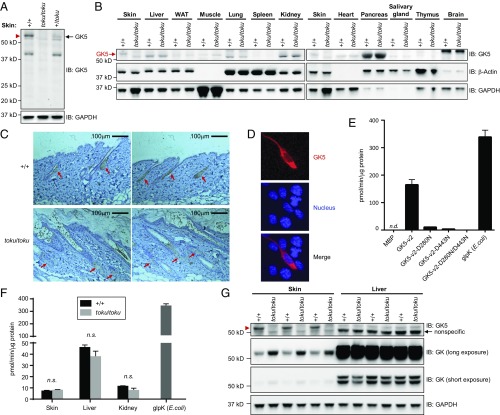
GK5 is predominantly expressed in the sebaceous glands of the skin and exhibits glycerol kinase activity. (A) Skin lysates from wild-type (+/+), toku heterozygote (+/toku), and toku homozygote (toku/toku) mice were immunoblotted using antibodies against GK5 and GAPDH. GAPDH was the loading control. (B) Lysates from skin, liver, white adipose tissue (WAT), muscle, lung, spleen, kidney, heart, pancreas, salivary gland, thymus, and brain from wild-type and toku homozygotes were immunoblotted using the indicated antibodies. β-Actin and GAPDH were the loading controls. (C) Immunohistochemical staining of P56 skin sections shows that GK5 is expressed mainly in the sebaceous glands in wild-type mouse skin; GK5 was not detected in the toku skin. Images were captured at 200× magnification. (Scale bar, 100 μm.) Representative images are shown. (D) FLAG-tagged GK5-v2 localized mainly in the cytoplasm in transfected NIH 3T3 cells. Immunofluorescence was performed using the FLAG antibody. DAPI stained the nuclei of the cells. All of the experiments were repeated at least three times, and a representative experiment is shown. (E) Glycerol kinase activity (pmol/min/μg) of recombinant wild-type GK5 or GK5 with one or both of the point mutations D280N and D443N. A coupling rate of 0.436 based on the manufacturer’s instruction was used to calculate the specific activities. Recombinant E. coli glycerol kinase (glpK; R&D Systems) is the positive control for the assay. Data represent means ± SD. (F) Total glycerol kinase activity (pmol/min/μg) in skin, liver, and kidney of wild-type and toku homozygotes. Recombinant glpK was used as a positive control. Data represent means ± SD. (G) Lysates from skin and liver from wild-type and toku homozygotes were immunoblotted using antibodies against GK5, GK, and GAPDH. GAPDH is the internal control.

