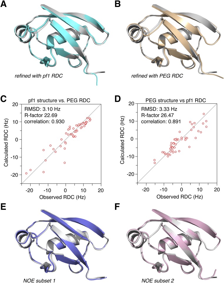
Fig. S6.
Cross-validation of the pUb retracted state structure. (A and B) Structures refined with only one set of the RDC restraints for backbone amide bond vectors, obtained in the alignment medium of either pf1 phage (cyan cartoon) or PEG(C12E5)/hexanol (yellow cartoon). Superimposed on the structure calculated with a full set of restraints (gray cartoon), the backbone RMS differences are 0.43 and 0.47 Å, respectively. (C and D) Correlations between the observed and calculated RDCs from the structure obtained with the RDC restraints excluded. The agreement is reasonably good for the cross-validated RDC restraints. (E and F) Structures refined with a subset of NOE distance restraints, with 10% of the NOEs randomly removed. The resulting structures have RMS differences of 0.29 and 0.27 Å from the structure calculated with the full set of restraints (gray cartoon), respectively.

