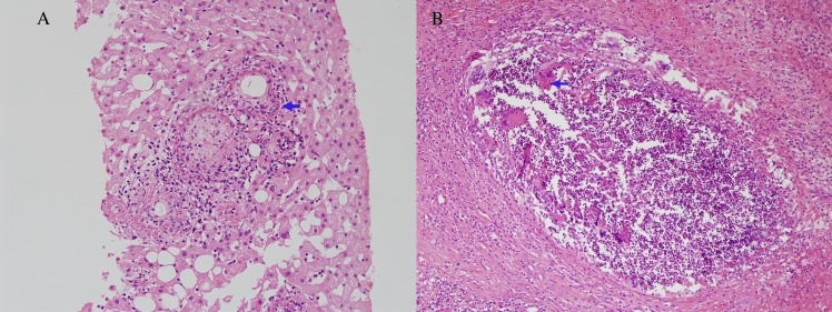
Fig 1. Representative photomicrographs of Q fever hepatitis (case no. 3 in case group, x200) and hepatic mucormycosis (case no. 6 in control group, x100).
(A) Characteristic fibrin ring granulomas consisting of a central fat globule or epitheloid cells with fibrin ring (arrow) (B) A suppurative granuloma consists of multinucleated giant cells with fungal hyphae (arrow) and polymorphous lymphoid cell including eosinophils.

