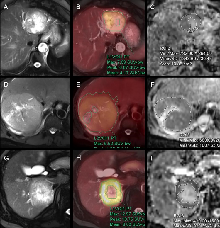
Fig 1. F-18 fluorodeoxyglucose (FDG) positron emission tomography (PET)/ magnetic resonance (MR) images with region of interest (ROI).
(A–C) FDG PET/MR images of 85-year-old woman with cholangiocarcinoma; (D–F) 46-year-old man with hepatocellularcarcinoma; (G–I) 72-year-old man with hepatic metastasis from colon cancer. (A, D and G) Axial HASTE MRI images; (B, E and H) Fused images of FDG PET and HASTE; (C, F and I) ADC map. ROIs were manually drawn along the contour of the tumor except necrosis. HASTE, half-Fourier acquisition single-shot turbo spin-echo; ADC, apparent diffusion coefficient.

