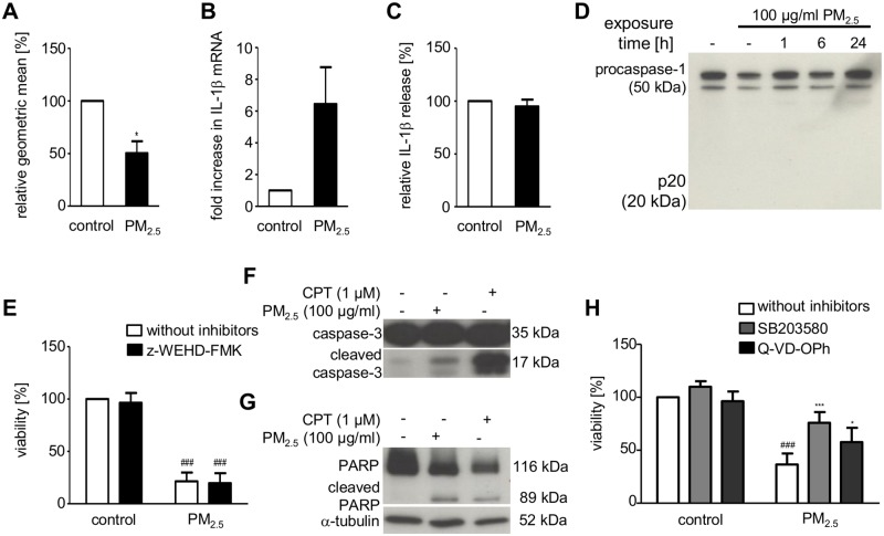
Fig 6. Effect of PM2.5 on lysosomal integrity, inflammasome activation, and apoptosis in BEAS-2B cells.
Cells were left untreated or exposed to 100 μg/ml PM2.5 one week before complete cell death. (A) Acridine orange staining and flow cytometry to determine lysosomal destabilization 72 h after re-exposure to PM2.5. Fluorescence intensity, displayed as the geometric mean of PM exposed BEAS-2B cells vs. untreated control cells (student’s t-test, *, p < 0.05; n = 3). (B) qRT-PCR for IL1β gene expression and (C) IL1β ELISA to quantify extracellular release of mature IL-1β 48 h after re-exposure to PM2.5 (student’s t-test, n = 3). (D) Immunodetection of procaspase-1 and its cleaved, activated fragment (p20) in whole cell lysates 0–24 h after re-exposure to PM2.5. (E) MTT cellular viability assay using the caspase-1 inhibitor z-WEHD-FMK (10 μM), added 1 h prior to re-exposure to PM2.5 for 72 h. Results show percentage of MTT conversion compared to untreated control cells (2-way ANOVA followed by the Bonferroni’s post-hoc test, ###, p < 0.001; n = 3). (F) Immunodetection of procaspase-3 processing to its active, cleaved form and (G) cleavage of the cellular caspase-3 substrate PARP in whole cellular lysates, prepared 24 h after re-exposure to PM2.5 (n = 3). Detection of α-tubulin served as a loading control. (H) MTT cellular viability assay using the p38 inhibitor SB203580 (10 μM) or the caspase-3/7 inhibitor Q-VD-OPh (10 μM), added 1 h prior to re-exposure to PM2.5 for 72 h. Statistically significant differences within groups are shown for BEAS-2B cells, left untreated or exposed to 100 μg/ml PM2.5 (2-way ANOVA followed by the Bonferroni’s post-hoc test, ###, p < 0.001) and PM2.5-exposed cells vs. PM2.5-exposed cells in the presence of SB203580 (2-way ANOVA followed by the Bonferroni’s post-hoc test, ***, p < 0.05) or Q-VD-OPh (2-way ANOVA followed by the Bonferroni’s post-hoc test, *, p < 0.001); n = 3.

