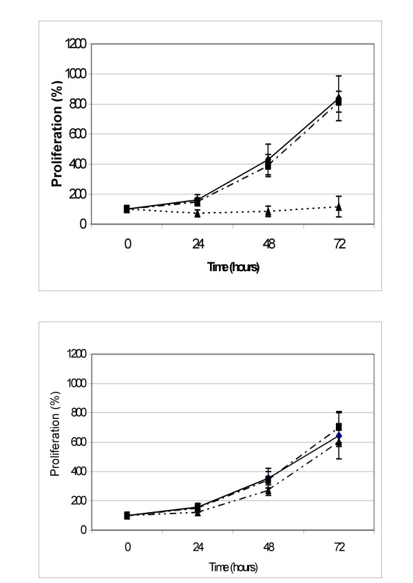
Figure 1.
K562wt cells (up) and K562adr cells (down) were incubated for 2 hours in medium alone (WT RPMI; full line and square, n = 6) supplemented in FCS 10% (dot line and lozenge, WT R10; n = 6), in methyl-β-cyclodextrin 5 mM (dot line and triangle, WT MCD; n = 6) and successively incubated in R10 for 72 hours. Data points are the percentages of the cellular concentration normalized to cellular concentration at T = 0 hours expressed as means, with vertical bars representing standard deviation (SD).

