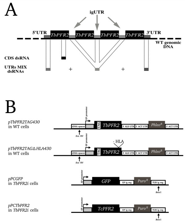
Figure 1.

dsRNAs and plasmids used for transfections. (A) Not-to-scale schematic representation of the endogenous TbPFR2 locus, with four copies of TbPFR2 coding sequence and specific UTRs. Regions targeted by RNAi are highlighted. The TbPFR2 coding sequence was targeted using CDS dsRNA and the UTRs were targeted all together with a set of dsRNA homologous to the 3' UTR, the intergenic UTR (igUTR) and the 5' UTR (UTRs MIX dsRNAs). (B) Not-to-scale representation of the constructs used for the transfection of WT cells (pTbPFR2TAG430 and pTbPFR2TAG430-ΔHLA; integration in the rDNA spacer) or TbPFR2i cells (pPCGFP and pPCTcPFR2; integration in the tubulin intergenic region). Large boxes represent protein coding sequences (black boxes: proteins of interest; grey boxes: antibiotic-resistance activities). Each plasmid was linearized with the indicated restriction enzyme prior to transfection into the cells. 3' ALD UTR: 3' UTR of the aldolase gene; ACT UTR: 5' or 3' UTR of the actin gene; EP ig reg: EP procyclin intergenic region; TUB ig reg: tubulin intergenic region.
