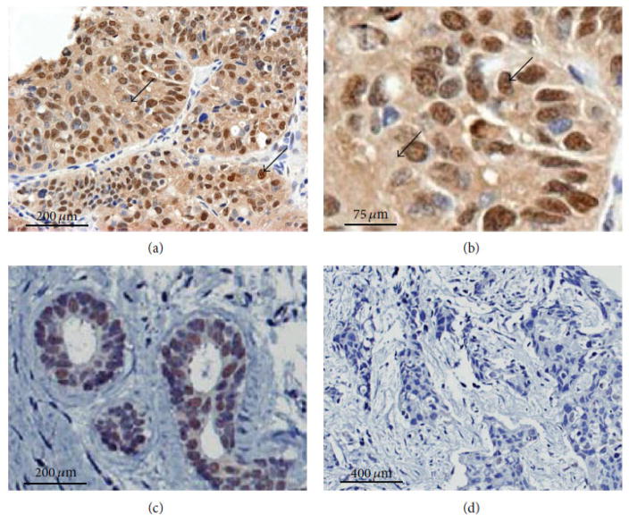
FIG. 1.
ERβ1 expression in archival TNBC specimens detected by immunohistochemisry using anti-ERβ1 antibody (PPG5/10, AbDSerotec). A representative example of ERβ1 immunostaining is shown using tumor and neighboring, nonmalignant tissue from the same patient. (a) Nuclear and cytoplasmic staining are shown on a representative specimen of TNBC at low magnification. (b) The same specimen from the previous panel shows nuclear and cytoplasmic staining at higher magnification as indicated by arrows. (c) Expression of nuclear ERβ1 is also present in neighboring normal tissue of the same patient tumor tissue displayed in panels (a) and (b). (d) A different TNBC tumor specimen that does not express ERβ1 is shown for comparison. Antibody binding was detected by using diaminobenzidine (DAKO). Sections were counterstained with Harris hematoxylin. (Reprinted with permission from the Hindawi Publishing Corporation, Copyright 2015).39

