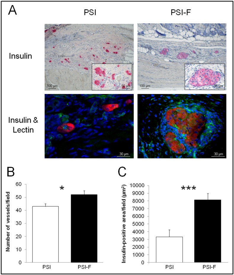
Fig 2. Histology of subcutaneous islet grafts 90 days after transplantation.
(A) Insulin immunostaining and insulin/lectin immunofluorescence (insulin in red to detect beta cells and lectin in green to detect vessels) performed on both PSI and PSI-F grafts. (B) Quantification of vessels in the subcutaneous grafts after CD31 immunostaining. (C) Quantification of the insulin-positive area in the subcutaneous grafts. *p<0.05; ***p<0.001.

