
Figure 1. Generation of H2O2 by DPEN and CuSO4 in breast and lung cancer cell cultures.
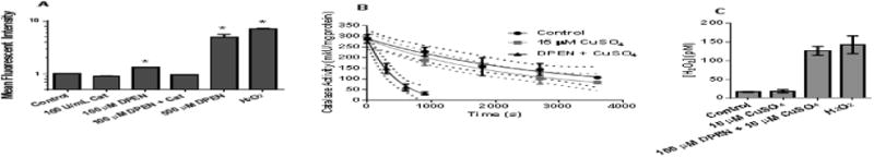
(A) MB231 cells were pre-incubated with PeroxyOrange-1 (PO-1) probe for one hour then treated with 100 μM DPEN, catalase (100 U/mL), or genuine H2O2 (100 μM) for 3 hours followed by analysis by flow cytometry. 15 μM CuSO4 was added to all groups at the time of drug treatment. *Significantly different than control; p< 0.05; n=4. (B) The rate of catalase inactivation in the presence of 3-aminotriazole was measured in H1299 lung cancer cells treated with 15 μM CuSO4 and 100 μM DPEN at various time points and (C) intracellular [H2O2] was calculated using the rates of catalase activity disappearance as previously described [23]. p<0.05; n=3. Errors represent ± 1SEM.