
Figure 1. Load-bearing periodontal ligament space (PDL-space), PDL-bone and PDL-cementum attachment sites, and PDL-inserts.
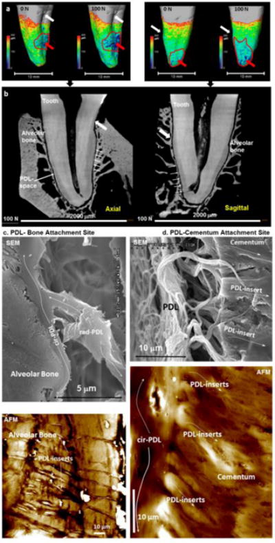
(a) Periodontal ligament (PDL)-space represented on the root surface of a human premolar at 0 N (mostly uniform PDL-space) and at 100 N (concentrated as pointed by the red arrow). Axial and sagittal virtual sections in two-dimensional (2D) space (b) are extrapolated from three-dimensional (3D) space (a). White arrows point to equivalent landmarks in both 3D (a) and 2D (b) spaces. Regions with significantly narrowed PDL-space specifically under loaded conditions are encircled in by a red curve (a) and corresponding regions in 2D space are indicated by arrows (b). The attachment of PDL to the alveolar bone (PDL-bone) and cementum (PDL-cementum) as visualized using scanning electron and atomic force microscopy (SEM, AFM) techniques is shown in panel c (PDL-bone) and d (PDL-cementum) respectively. Note a change in radial-PDL (rad-PDL) orientation to circumferential PDL (cir-PDL) as it “skirts” along alveolar bone and cementum before it inserts (PDL-inserts) into respective mineralized tissues within the load-bearing complex. In both cases these inserts are hygroscopic as denoted by their increased height profile when imaged under hydrated conditions (white regions correlate to topographical peaks of 2000–3000 nm compared to darker regions, which correlate to 0 nm).