
Figure 2. Physical characteristics of a normal and compromised PDL and the bone-PDL-cementum complex using various high resolution imaging and AFM-based nanoindentation techniques.
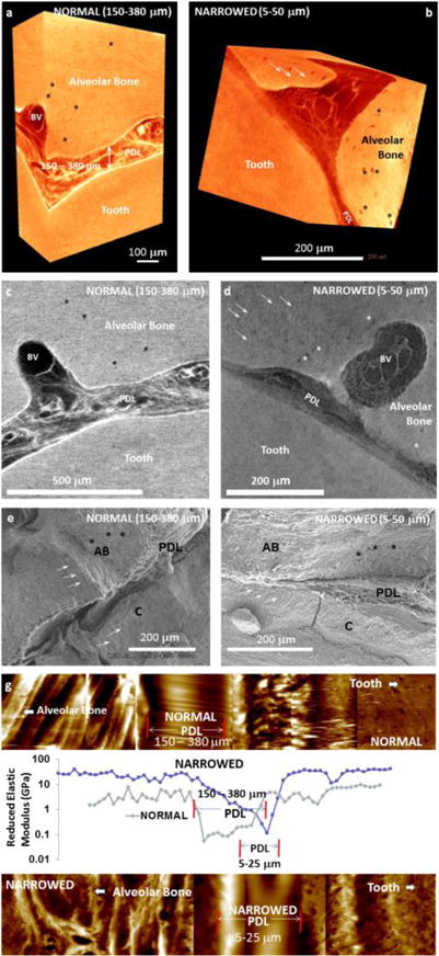
Digitally segmented blocks from human molars illustrate the PDL under seemingly normal (a) and compromised conditions (b). Normal condition is identified as a PDL-space of 150–380 μm (a), and the narrowed PDL-space illustrates 5–50 μm (b). Similar to the 3D digital reconstructions, virtual sections illustrate vascular bundles (blood vessels: BV) within the normal PDL, and similar structures are visible in narrowed PDL (d). Additionally, PDL-inserts can be observed in less X-ray attenuating regions in 3D reconstructed volumes and 2D virtual sections (white arrows in b and d). Similarly osteocytic lacunae are also identified (asterisks in a-d). Scanning electron micrographs of normal (e) and narrowed (f) conditions illustrate PDL between alveolar bone (AB) and cementum (C). Elastic modulus mapping under wet conditions using a nanoindenter illustrate a discontinuity in the narrowed bone-PDL-cementum complex (g).