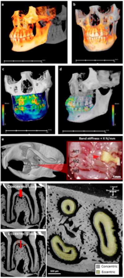
Figure 3. Clinical scenario that exploits manipulating PDL to promote orthodontic tooth movement, and an animal model to link physical perturbations on the crown to biological processes in the bone-PDL-tooth load-bearing fibrous joint.

Orthodontic intervention is a clinical scenario that could stimulate plausible discontinuities in physical characteristics of the bone-PDL-tooth complex. Overall tooth displacement by comparing cone-beam computed tomographic reconstructed volumes taken before and after one year of orthodontic intervention is illustrated. Note that observed maximum tooth-displacement is 4 mm and is significantly larger than the 150–380 μm of PDL-space generally identified in humans. Physical changes due to mechanical stimulation resulting from biological processes to prompt tooth translation are programmed in rodents, specifically in mice between the molars 1 and 2 (M1 and M2, red arrow head) with an elastic band of known stiffness (X N/m) (e) to first perform comparative analyses on PDL-space changes (h) under centric (f) and eccentric (g) conditions. Note that in the illustrated scenario the roots of an eccentrically loaded first molar were displaced in the distal direction (h) specifically when compared concentric conditions (g).
