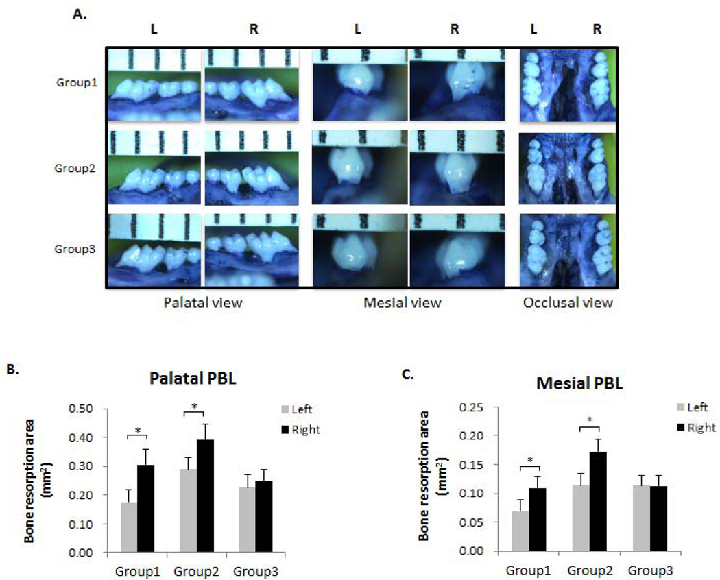
Figure 2. OF increased PBL of ligature-induced experimental periodontitis.
(A) Palatal, mesial and occlusal view of the de-fleshed maxilla stained with 1% toluidine blue solution. Maxillary images were captured with a digital stereomicroscope system on a holder to enable the visualization of the cementoenamel junction (CEJ) and alveolar bone crest. The polygonal areas palatal and mesial to the first molars were measured using Image J (NIH) software. L, left side (represents control side in Group 1 and SL side in Groups 2&3); R, right side (represents SL side in Group 1 and SL+OF side in Groups 2&3). n = 6–8. (B) Morphometric analysis of bone resorption area palatal to the maxillary first molars. (C) Morphometric analysis of bone resorption area mesial to the maxillary first molars. Bone resorption areas were significantly increased on the right side compared to the left side. *p < 0.05. n = 6–8.

