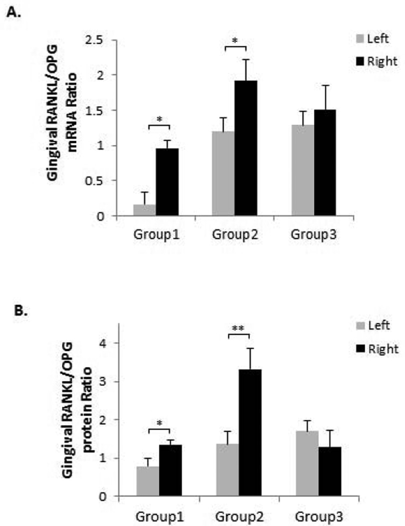
Figure 3. OF increased gingival RANKL/OPG expression ratio.
(A) Gingival mRNA expressions of RANKL and OPG were detected by real-time PCR and their ratios were calculated. RANKL/OPG ratios were increased on the right side compared to the left side in Groups 1 and 2. For all the experiments, GAPDH gene was used as internal control. (B) Gingival protein levels of RANKL and OPG were detected by ELISA and their ratios were calculated. RANKL/OPG protein ratios were also increased on the right side compared to the left side in Groups 1 and 2. *p < 0.05, **p < 0.01. n = 6–8.

