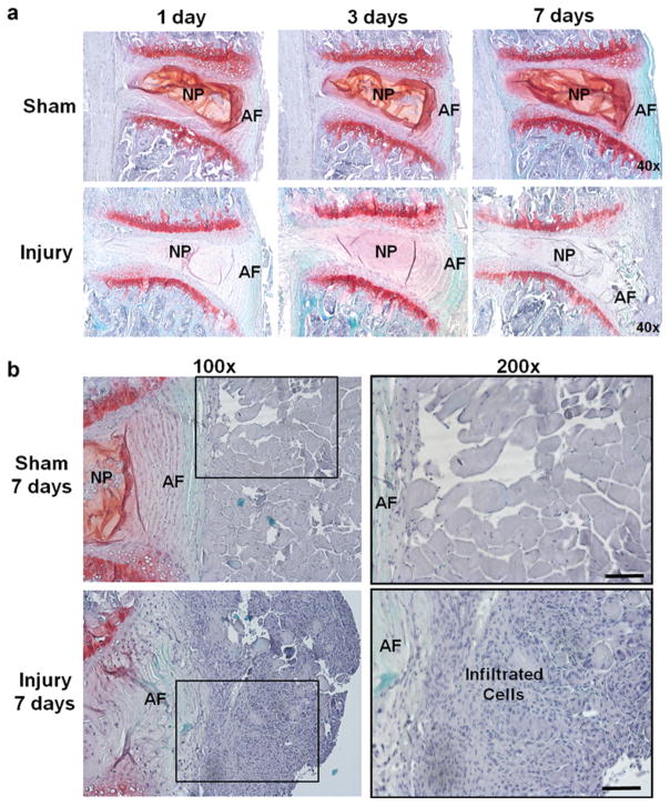
Fig. 1.
Safranin-O staining of injury and sham disks in WT mice. a At various time points (1, 3, and 7 days) after surgery, punctured disks demonstrated structural change and decreased proteoglycan content confirming disk herniation induced by the needle puncture (×40). b Higher magnification images of safranin-O staining illustrated significant amount of inflammatory cell infiltration in the region adjacent to punctured AF in injured disks at 7 days after surgery (lower row), whereas no such cell infiltration was found in the sham group (upper row). The black square in the left column (×100) suggested tissue area magnified in the right column (×200) (scale bar = 100 μm).

