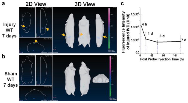
Fig. 4.
Representative in vivo NIRF imaging with cFLFLF-PEG-Cy7 at 7 days post probe injection. a Strong fluorescence signal was identified around injured disks on lumbar spines of WT mice, whereas b no signal was detectable in the corresponding lumbar region of sham. c In vivo observed fluorescence intensity of disk injury region was plotted against post probe injection time (n = 5).

