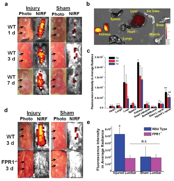
Fig. 5.
Ex vivo NIRF disk imaging of WT and FPR1−/− mice with cFLFLF-PEG-Cy7. a Representative photographs and NIRF images of WT lumbar spines containing both injured and intact disks at various time points post probe injection. b Organ distribution profile of cFLFLF-PEG-Cy7 at 7 days post probe injection. c Epi-fluorescence intensity of various organs, tissues, and punctured and sham lumbar spines at 1, 3, and 7 days post probe injection (n = 5 per group). *p = 0.0084 compared to sham lumbar at 3 days, **p = 0.0027 compared to sham lumbar at 1 and 7 days, respectively. d Representative fluorescence imaging of WT and FPR1−/− lumbar spine containing punctured and intact disks at 3 days post probe injection (images were displayed under the same color scale). e Fluorescence intensity of injury and sham lumbar for wild-type and FPR1−/− mice (n = 5 per group). For WT/injury group, *p = 0.0084 vs. WT/sham, *p = 0.0035 vs. sham/FPR−/−, *p = 0.0029 vs. injury/FPR−/− compared to all other groups. No statistically significant difference was observed among sham/WT, injury/FPR1−/−, and sham/FPR1−/− groups (p = 0.85 for injury/FPR1−/− vs. sham/FPR1−/−, p = 0.75 for injury/FPR1−/− vs. sham/WT).

