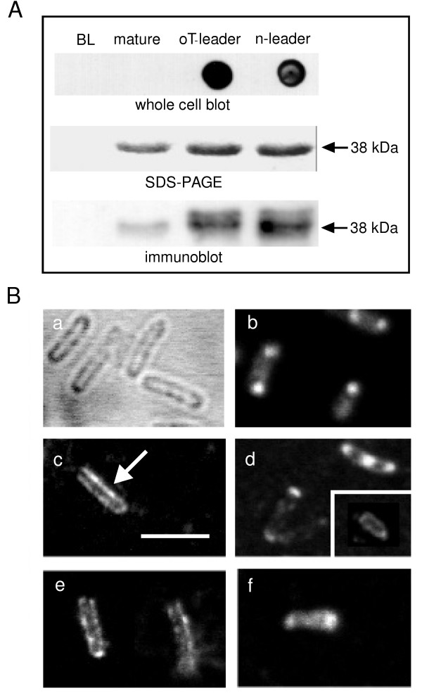
Figure 3.

Insertion of MOMP into the E. coli outer membrane. A. Recombinant C. trachomatis MOMP was expressed for 2 hrs at 25°C from constructs encoding either no leader (mature), the OmpT (oT) leader, or the native (n) leader, and immunodetected on the surface of intact BL21 cells (upper panel) using a specific anti-MOMP polyclonal Ab. Non-expressing BL21 cells (BL) show no signal, and mature, leaderless MOMP does not reach the cell surface. The middle and lower panels show, respectively, SDS-PAGE and immunoblot analyses of the corresponding recombinant proteins under these conditions (prepared as in Fig. 2). Note the presence of some unprocessed protein, revealed by the immunoblots of leadered protein expression. Representative of 3 similar experiments. B. Immunofluorescence confocal microscopy (panels b–f), with examples of unstained cells (panel a); cells permeabilised and stained after expressing mature, non-leadered MOMP (panel b); cells expressing OmpT-leadered MOMP stained before (panel c) and after permeabilisation (panel d, with inset permeabilised omp8 cell after 12 hrs induction at 16°C); cells expressing native-leadered MOMP under corresponding conditions (panels e and f, respectively). The scale bar (panel c) is 3 μm, and the arrow points out membrane staining. Representative of 3 similar experiments.
