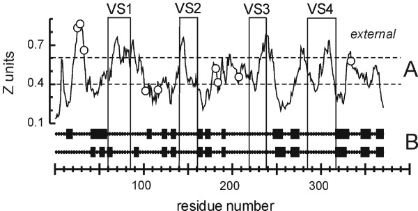
Figure 4.

Membrane topology and secondary structure predictions for C. trachomatis MOMP. A. "Membrane crossing" prediction. Surface-exposed VS domains and cysteine residues are indicated by boxes and circles, respectively. A "complete membrane crossing" corresponds to a (contiguous) region of the plot that crosses both dotted lines in sequence, where the dotted lines represent the internal (periplasmic) and external borders of the outer membrane (in Z units). B. Two independent β-strand predictions, TMBETA [38] (upper line) and B2TMPRED [37] (below). The strands are boxed. The residue numbers refer to the mature (processed) protein.
