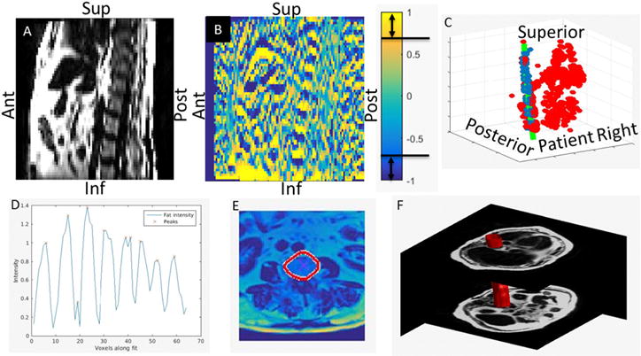
Figure 1.

Sagittal plane of a fat image (A) shown side by side with the axial gradient of the same section (B). Gradient voxels containing directional cosine magnitudes greater than 0.7 are considered candidate vertebral body edges. The collection of candidate voxels (all points in (C)) are fit by a 3D quadratic shape model using RANSAC (green line in (C)). In panel (C), the inliers are colored blue and the outliers are colored red. The maxima of the fat image are then identified along the curve fit (D). These positions are then used as starting points for an active contour algorithm to expand to the edges of the vertebral bodies (E). The vertebral body envelope is created by interpolating each of the vertebral body contours in the superior–inferior direction (F).
