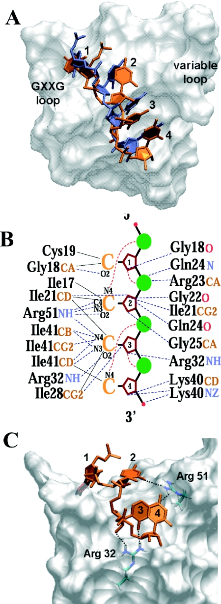
Figure 5.
(A) Molecular surface of αCP1-KH3 showing modelled position of poly(C)-RNA (orange) based on the Nova2-KH3-RNA structure (accession no. 1EC6). The poly(C)-tetrad is viewed from above the GXXG and variable loops, highlighting their position either side of the hydrophobic binding cleft. (B) Summary of potential interactions occurring between the modelled αCP1-KH3 and poly(C)-RNA. A poly(C)-RNA-tetrad is represented schematically. Potential hydrogen bond interactions are indicated by dotted lines. Those from specific residue atoms to the RNA backbone are listed on the right, and those to the cytosine bases are listed on the left. The red dotted lines represent intra-molecular hydrogen bonds that may stabilize the RNA in its binding mode to the KH domain. Hydrophobic or van der Waals contacts to the cytosine bases are indicated by solid lines. (C) The positions of Arg 32 and Arg 51 side chains are highlighted beneath the molecular surface of αCP1-KH3. Potential hydrogen bonds to the poly(C)-RNA are shown as dotted lines.

