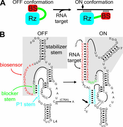
Figure 1.
The concept of the SOFA-ribozyme. (A) Schematic representation of both the ‘off’ and ‘on’ conformations of the SOFA-ribozyme. The ribozyme (Rz) and the biosensor (BS) are in blue and red, respectively. The small arrow indicates the cleavage site. (B) Secondary structure and nucleotide sequence of SOFA+-δRz-303 in both the ‘off’ and the ‘on’ conformations. The gray section indicates the SOFA module. The P1 stem of the ribozyme and the biosensor are in blue and red, respectively. The blocker sequence is represented in green. The C76A mutation that produces an inactive ribozyme version is indicated. The numbering system is according to (15).

